The Radiology Assistant Bowel wall thickening CTpattern
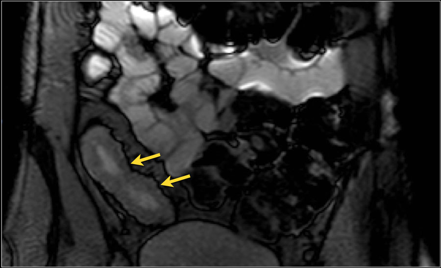
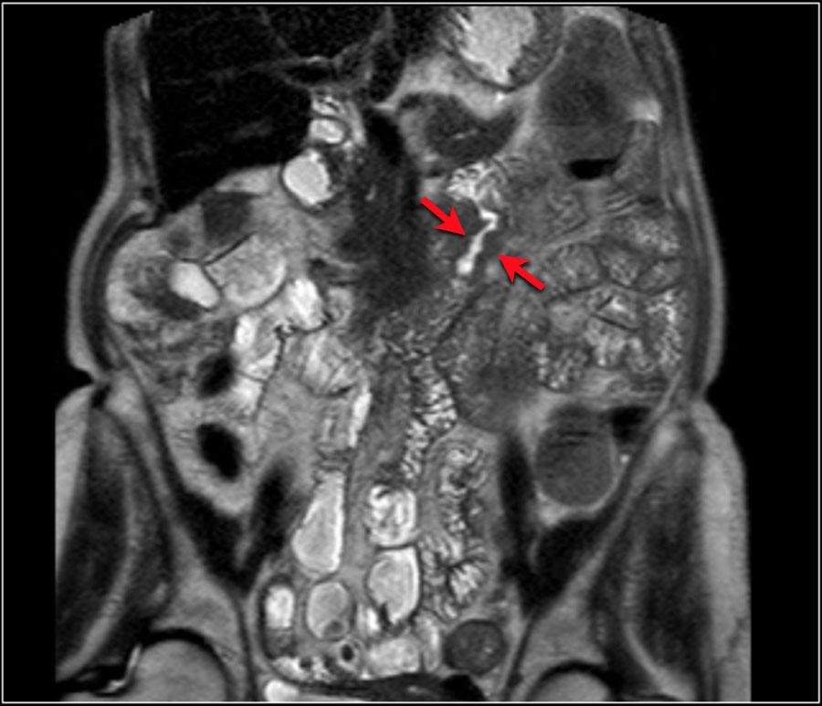
A perianal fistula is an abnormal connection between the epithilialised surface of the anal canal and the skin. Obstruction of anal gland which leads to stasis and infection with absces and fistula formation (most common cause). Inflammatory bowel diseases (Crohn's disease more common than colitis ulcerosa)
INFLAMMATORY BOWEL DISEASE IMAGING(RADIOLOGY)
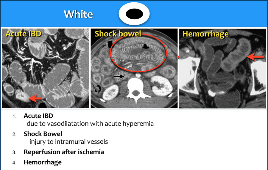
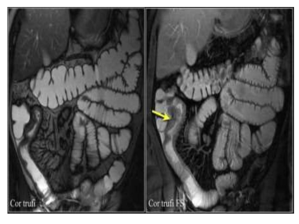
The current status of MRI in ulcerative colitis is that of a promising, non-invasive technique for imaging extent of more severe disease. The most striking abnormalities in ulcerative colitis are colonic wall thickening and increased enhancement. The median wall thickness in ulcerative colitis ranges from 4.7 to 9.8 mm.

Inflammatory Bowel Disease Body MRI
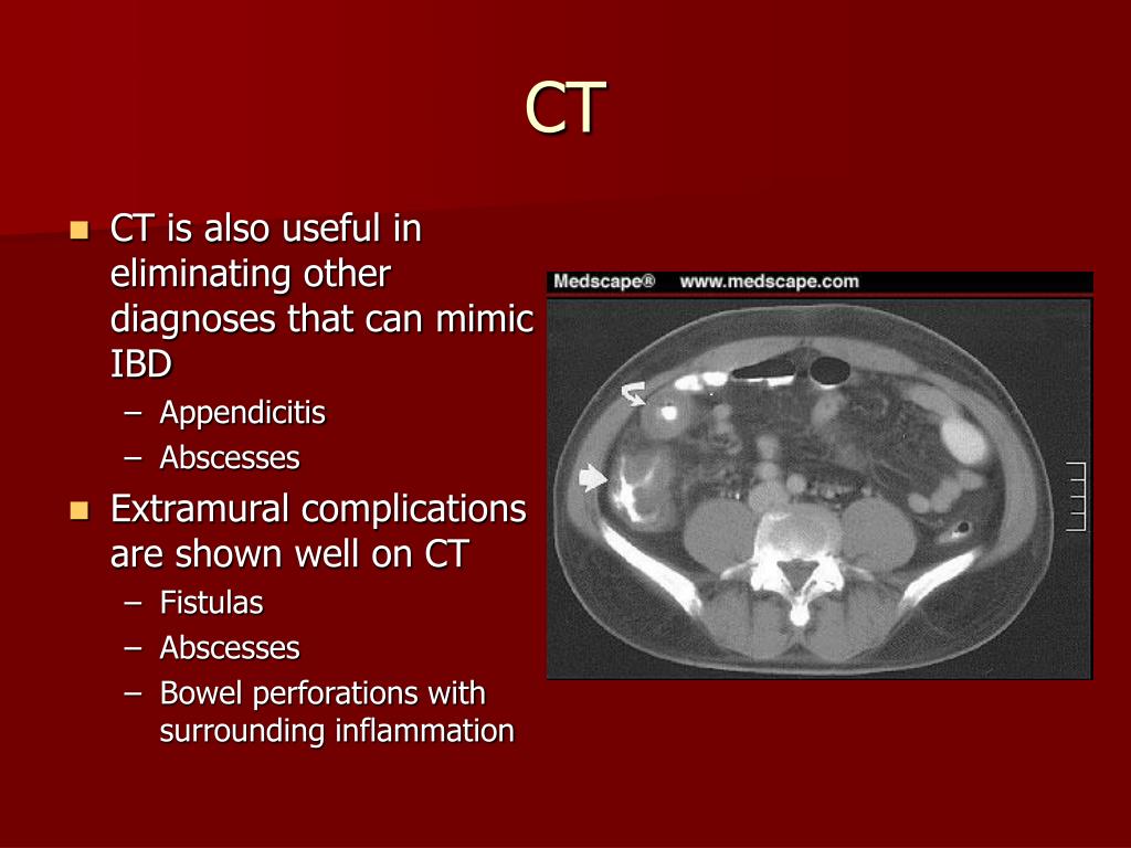
The purpose of this paper is to evaluate the role of imaging in inflammatory bowel disease (IBD), including detection of extraluminal complications and extraintestinal manifestations of IBD, assessment of disease activity and treatment response, and discrimination of inflammatory from fibrotic strictures.. Radiology. 2006; 238:517-530.
The Radiology Assistant Crohn's disease
Inflammatory bowel disease (IBD), although occasionally used to encompass a variety of infective and purely inflammatory bowel conditions, usually refers to two idiopathic conditions: Crohn disease ulcerative colitis Indeterminate colitis is added to the list and represents ~6% of inflammatory bowel disease cases 2.
The Radiology Assistant Small Bowel Tumors
Technical considerations MRI of the bowel (like abdominal MRI in general) is based on the principles of ultra-fast imaging. In the past, most examinations were performed on 1.5T scanners equipped with strong gradient systems. Only recently, bowel MRI on 3T systems has proved feasible. 19

Computed Tomography Enterography and Inflammatory Bowel Disease Abdominal Key
This work makes recommendations for imaging findings that indicate small bowel Crohn's disease, how inflammatory small bowel Crohn's disease and its complications should be described, elucidates potential extra-enteric findings that may be seen at imaging, and recommends that cross-sectional enterography should be performed at diagnosis of Crohn.
Thickened Small Bowel Loops due to Inflammatory Bowel Disease (IBD) Small Bowel Case Studies
Radiology Crohn's Disease-Focused Panel, the Society of Pediatric Radiology, the American Gastroenterological Association, and other experts, systematically evaluated evidence for imaging findings associated with small bowel Crohn's disease enteric inflammation and established recommendations for the evaluation, interpretation, and
Inflammatory Bowel Disease Small Bowel Case Studies CTisus CT Scanning
Abstract This online presentation describes a didactic way to learn the distinct and overlapping clinical, radiologic, and pathologic characteristics of the two major conditions related to inflammatory bowel disease: ulcerative colitis and Crohn disease. Article History Received: Mar 7 2016 Revision requested: May 16 2016
PPT Radiographic Imaging in Inflammatory Bowel Disease PowerPoint Presentation ID6712333
This article is based on a presentation given by Richad Gore and adapted for the Radiology Assistant by Robin Smithuis. Richard Gore is editor of the Textbook of Gastrointestinal Radiology, 3rd Edition and High Yield Imaging: Gastrointestinal.

INFLAMMATORY BOWEL DISEASE Radiological interpretation for Doctors, Medical students, Nurses
The use of cross-sectional imaging and ultrasonography has long complemented endoscopic assessment of inflammatory bowel disease (IBD). Clinical symptoms alone are often not enough to assess disease activity, so a reliance on non-invasive techniques is essential. In this paper, we aim to examine the current use of radiological modalities in aiding the management of patients with IBD. We focus.
INFLAMMATORY BOWEL DISEASE IMAGING(RADIOLOGY)
Sensitivity of MRI for the number of affected bowel segments was 97.5% with a specificity of 100%, whereas ultrasound scored 76% and 75%, respectively. For stenosis, MRI had a sensitivity of 100% and a specificity of 96%, and ultrasound a sensitivity of 58% and specificity of 100%.
Inflammatory Bowel Disease Small Bowel Case Studies CTisus CT Scanning
The current biomarkers used in IBD monitoring are C-reactive protein (CRP) and fecal calprotectin. Based on data from the CALM study, a combination of CRP and fecal calprotectin most accurately predicts relapse ( Lancet 2017;390 [10114]:2779-2789 ). "The CALM study showed that CRP and fecal calprotectin biomarker levels can guide treatment.

INFLAMMATORY BOWEL DISEASE IMAGING(RADIOLOGY)
Endoscopic assessment of inflammatory bowel disease (IBD) can often be imprac-tical and burdensome on both patient and clinician. Complementing endoscopy with radiological imaging has long been established practice in IBD care. With advancements in imaging, there have been great strides in the disease moni-toring and management of IBD. Different
Inflammatory Bowel Disease Colon Case Studies CTisus CT Scanning
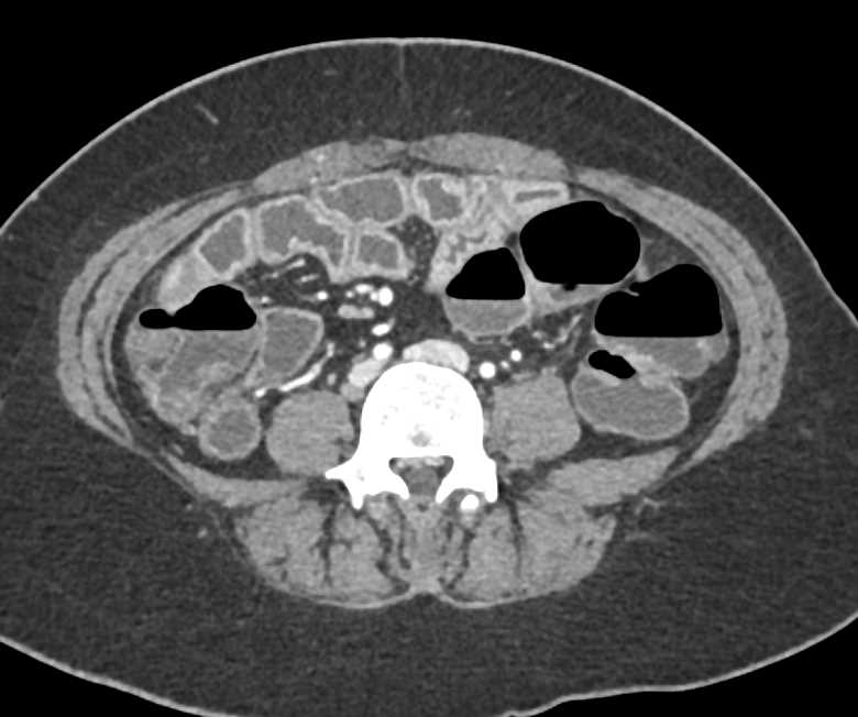
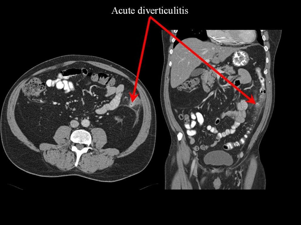
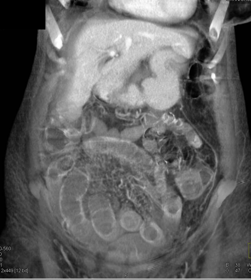
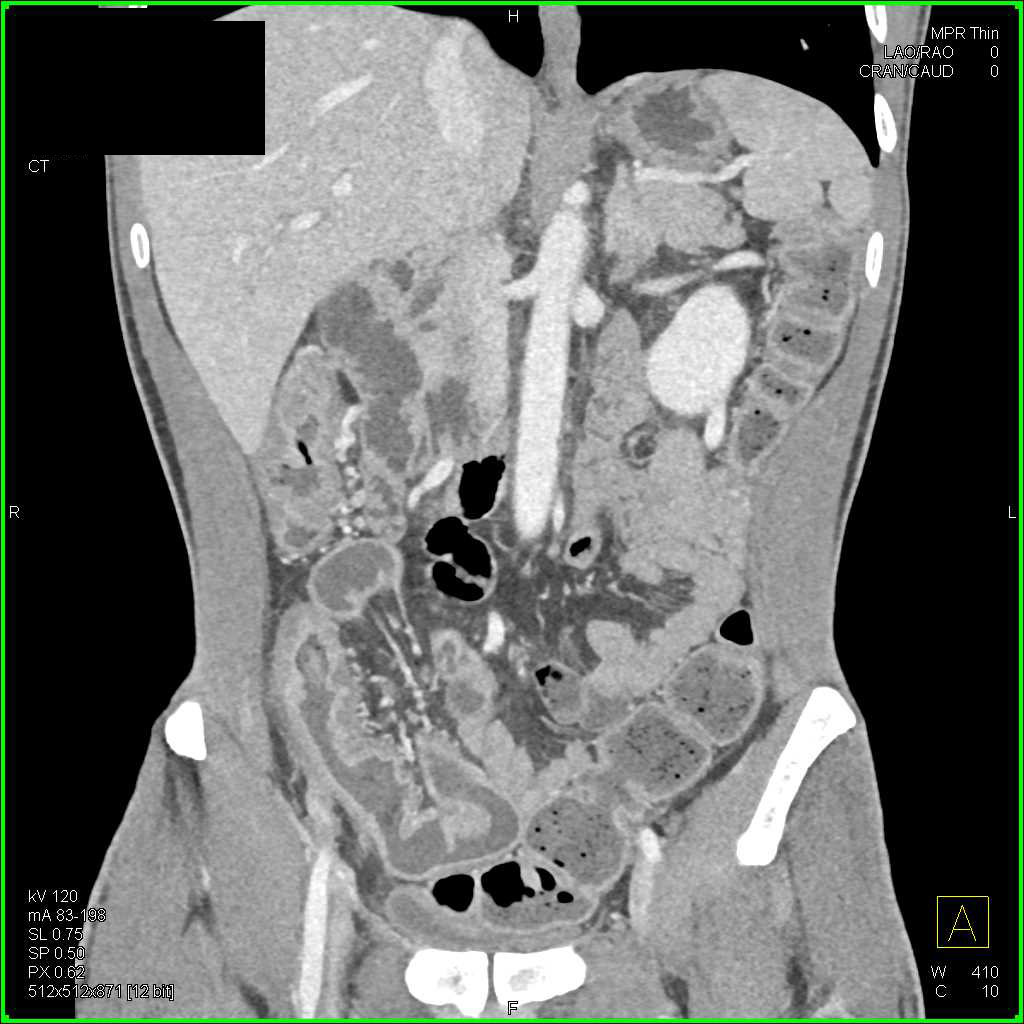
Crohn's disease is characterized by inflammatory lesions in the gastrointestinal tract, most commonly in the terminal ileum and colon. The lesions are usually transmural, which can lead to complications like stenoses, fistulas and abscesses.
INFLAMMATORY BOWEL DISEASE IMAGING(RADIOLOGY)
Crohn disease, also known as regional enteritis, is an idiopathic inflammatory bowel disease characterized by widespread discontinuous gastrointestinal tract inflammation. The terminal ileum and proximal colon are most often affected. Extraintestinal disease is common. Epidemiology

Inflammatory Bowel Disease Current Role of Imaging in Diagnosis and Detection of Complications
This article, authored by the Society of Abdominal Radiology Crohn's Disease-Focused Panel, illustrates the imaging findings and recommended radiology report impression statements described in the consensus recommendations with examples of CT enterography and MR enterography images.